Skip to content
Julie AupetitUltrasound midwife(back to home page)

Ultrasound scans

Scans performed at the practice

Obstetric and gynaecological ultrasound at the Pôle Santé in Juvignac. Select a scan to learn more.

Choose a scan

Dating

Pregnancy follow-up

Gynaecology & fertility

Dating scan

The dating scan, usually performed between 6 and 8 weeks of amenorrhoea (counted from the first day of your last period), confirms that the pregnancy is inside the uterus and progressing.

For an unwanted pregnancy

The dating scan is the examination required before a termination of pregnancy.

The scan follows the code of good practice drawn up by the Occitanie Perinatal Network. You will be welcomed with kindness and discretion. No image and no sound will be imposed on you; the images taken during the scan are stored in your secure record and are not handed over unless you ask for them.

You will receive a report with the information needed by the healthcare professional who will see you for the next steps of your care.
For a wanted pregnancy

Depending on the stage of pregnancy, it shows the embryo and its heartbeat, which indicates that the pregnancy is progressing.

Measuring the embryo gives a first estimate of the date the pregnancy began, before the measurement taken at the first-trimester scan.

Pregnancy screening scans

Need to work out your scan dates?Date calculator (Gestaclic, in French) (opens in a new tab)

How does a pregnancy scan work?

  1. 1Ultrasound uses sound waves to produce images of the baby. It does not use X-rays.
  2. 2You will usually lie on your back or slightly on your side. Gel is applied to your belly, then a probe is moved over the skin to obtain the images. The scan is usually painless, although you may sometimes feel slight pressure on your belly.
  3. 3The length of the scan can vary depending on the baby’s position, the scanning conditions and what needs to be examined.
  4. 4Please bring the results of any previous tests or scans, if you have them.

Your consent and your questions

You can ask the professional performing the scan any questions you have. Screening is different from diagnosis: when an anomaly is suspected, further tests may be needed to confirm or rule it out.

The aim is to give you clear information so that you can understand the scans offered and their results.

A question before your appointment? Message me through Doctolib.

A normal scan does not guarantee that “everything is fine”

Screening scans are offered to inform you about how your pregnancy is progressing and to look for certain anomalies, following precise national guidelines and required images (CNEOF 2023 report, in French (opens in a new tab)).

Even when a scan is normal, it cannot rule out every anomaly, as some may appear later in pregnancy or after birth.

If an unusual image or a scanning difficulty is noted, it does not necessarily mean the baby has an anomaly. An additional scan may simply be offered to obtain more precise images.

1

First scan: between 11 and 13 weeks + 6 days

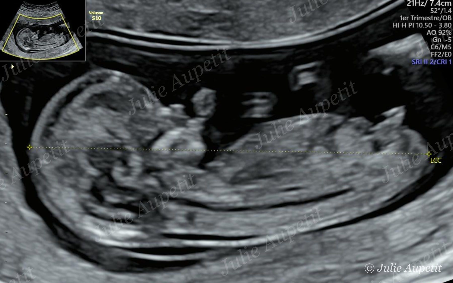
It dates the pregnancy and confirms the due date, checks the number of babies and the baby’s general development, and measures the nuchal translucency (a small area at the back of the baby’s neck). Nuchal translucency measurement contributes to screening for certain chromosomal anomalies, especially trisomy 21 (Down syndrome). On its own, it cannot provide a diagnosis.

First-trimester ultrasound: baby in profile with crown-rump length measurement
2

Second scan: around 22 weeks

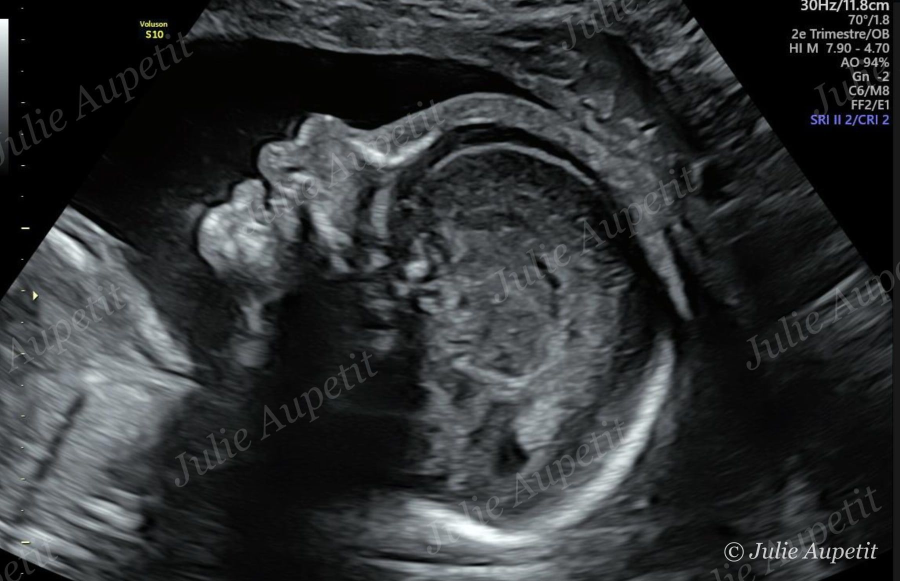
This is the anomaly (morphology) scan. The professional examines different parts of the baby in detail, as well as growth, the placenta and the amniotic fluid. This scan looks for certain malformations that may be visible at this stage of pregnancy.

Second-trimester anomaly scan: baby’s profile
3

Third scan: around 32 weeks

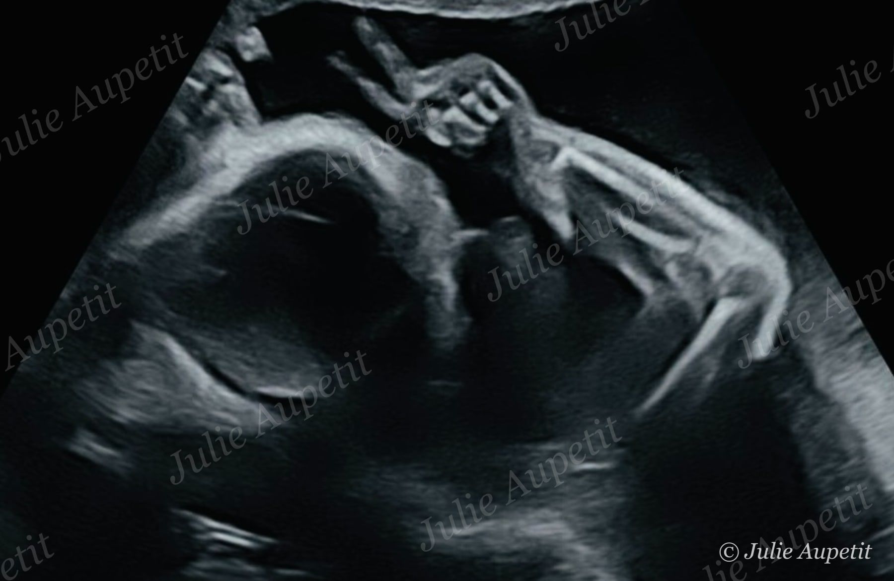
It checks the baby’s growth and development, the placenta and the amniotic fluid. It also looks for certain anomalies that may only become visible later in pregnancy.

Third-trimester ultrasound: baby with a hand near the face

Gynaecological ultrasound

Pelvic ultrasound

A pelvic ultrasound may be performed in various situations: pelvic pain, unusual bleeding, menstrual problems, gynaecological check-up, monitoring of a cyst or fibroid, infertility, or to look for the cause of certain symptoms.

How is the scan performed?

Depending on the situation, the scan can be performed in two ways.

Transabdominal

A probe is moved over your lower abdomen after applying gel. You may be asked to have a full bladder to improve image quality, especially if you have never had sexual intercourse, as the transvaginal route is then not appropriate.

Transvaginal

A thin probe, covered with a single-use protective sheath and gel, is gently inserted into the vagina. This painless technique provides more precise images of the uterus and ovaries.

Can the scan be performed during my period?

Yes. Periods do not usually prevent the scan. However, depending on the reason for the scan, some phases of the cycle may be better suited to certain measurements or observations: feel free to contact me for more information.

The professional will explain the results and tell you, if necessary, whether follow-up or an additional examination is recommended.

Fertility ultrasound and ovulation monitoring

Pelvic ultrasound is an important part of infertility assessment and ovulation monitoring.

This scan is prescribed by your healthcare provider as part of assisted reproduction (ART) care. Ovulation monitoring takes place during ovarian stimulation.

It allows the follicles to be counted and measured, the thickness of the endometrium to be assessed, and the blood flow to your uterus to be studied. Depending on the situation, the scan may be combined with hormonal blood tests and/or ovulation tests.

Ready for your scan?

Appointments can be booked online on Doctolib.